Versatile research tool unlocks galectin-1 biology across pre-clinical assay workflows
HUSTON, TX, UNITED STATES, September 16, 2026 /EINPresswire.com/ — Understanding glycan-mediated cell-to-cell communication, widely known as the biological “sugar code”, represents one of the most compelling frontiers in modern life-science research. Galectin-1 (GAL1), a multifunctional glycan-binding protein, governs critical immune homeostatic and oncogenic signalling pathways. Reliable antibody reagents are indispensable for deciphering its dual biological roles. Cloud-Clone presents validated GAL1 antibody that delivers consistent performance across a full spectrum of laboratory applications, supporting global researchers to advance immunology and tumour biology investigations.
On the microscopic stage of life science, proteins act as central performers. Highly orchestrated interactions between these molecules underpin all ordered biological activities. This article centres on Galectin-1 (GAL1), a unique protein with dual biological roles as both a physiological protector and a tumour-promoting mediator, and introduces GAL1 antibody, a critical research reagent enabling scientists to unravel GAL1-driven biological mechanisms.
GAL1: A Core Protein for Deciphering the “Sugar Code”
GAL1 is a β-galactoside-binding lectin and the earliest-discovered member of the galectin protein family. Despite its small molecular weight of approximately 14.5 kDa, it exerts extensive biological functions. Functioning as a high-precision molecular key, GAL1 specifically recognises and binds complex cell-surface glycan chains carrying β-galactoside residues. This molecular recognition process is metaphorically described as reading the cellular “sugar code”.
Thanks to this distinctive glycan-recognition capacity, GAL1 participates in broad molecular crosstalk within human organisms and drives divergent biological outcomes.
A Double-edged Sword Within the Immune System
Numerous studies have confirmed that GAL1 acts as a double-edged sword in immune-related biological processes.
Homeostasis-maintaining factor under physiological conditions Under normal physiological circumstances, GAL1 serves as a key modulator sustaining immune homeostasis. It induces apoptosis of activated effector T-cells, putting timely brakes on excessive immune responses. Such activity is essential for restraining inflammatory responses and preventing autoimmune disorders. GAL1 also facilitates clearance of exhausted immune cells to maintain a balanced immune microenvironment.
Tumour-promoting mediator under pathological conditions During tumour progression, GAL1 undergoes a dramatic functional shift. Tumour cells and stromal cells within the tumour microenvironment (TME) secrete large quantities of GAL1, which becomes a potent mediator enabling tumour immune escape via multiple mechanisms:
– Direct elimination of immune cells: GAL1 binds and cross-links glycosylated receptors such as CD43 and CD45 expressed on activated T-cells, triggering apoptosis in both CD4+ and CD8+ effector T-cells.
– Induction of immune-suppressive cell populations: GAL1 drives proliferation and differentiation of regulatory T-cells (Treg) and reprograms dendritic cells (DC) toward a tolerogenic phenotype, constructing a microenvironment favourable for tumour survival.
– Remodelling of the tumour microenvironment: GAL1 promotes M2-type pro-tumour macrophage polarisation and interacts with molecules including VEGFR2 to stimulate tumour angiogenesis, supplying nutrients and metastatic routes for tumour lesions.
Accordingly, GAL1 expression correlates with poor clinical prognosis across multiple cancer types. In non-small-cell lung cancer (NSCLC), elevated GAL1 plasma levels have been documented. Patients with high GAL1 expression exhibit shortened median progression-free survival following anti-PD-1 immunotherapy, indicating that GAL1 may dampen immunotherapeutic efficacy. In liver cancer, GAL1 is recognised as one of the key drivers that propel disease progression from hepatitis and fibrosis toward malignant transformation. These observations establish GAL1 as a high-potential therapeutic target and prognostic biomarker for oncology research.
GAL1 Antibody: A Multifunctional Research Reagent
High-specificity, high-sensitivity detection tools are required to dissect GAL1’s complex activities and profile its expression patterns under physiological and pathological conditions. GAL1 antibody stands out as a core research reagent. Both polyclonal antibody (pAb) and monoclonal antibody (mAb) formats can selectively recognise and attach to GAL1 protein, functioning like precision-guided molecular probes.
Distinct experimental objectives call for different assay applications of GAL1 antibody:
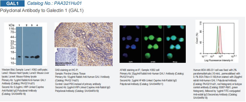
Western Blot (WB): Protein identity verification At a working concentration of 1 µg/mL, GAL1 antibody accurately detects GAL1 protein within complex cell or tissue lysate samples including K562 cells and mouse heart, liver and kidney tissues. Researchers can verify target molecular weight and compare relative protein expression levels. WB represents the fundamental technique for profiling GAL1 protein abundance.
Immunohistochemistry (IHC): Spatial localisation within tissues For IHC assays performed at 10 µg/mL working concentration, the antibody successfully stains GAL1 within porcine uterine tissue and visualises its spatial distribution across tissue sections. Negative control samples using PBS in place of primary antibody yield no detectable staining signal, confirming high binding specificity. This assay type helps researchers identify cellular sources and positional patterns of GAL1 within tissues or tumour microenvironments.
Immunofluorescence (IF): Subcellular visualisation via microscopy Immunofluorescence imaging reveals intracellular localisation of GAL1. Tested on K562 cells at 20 µg/mL working concentration, GAL1 antibody paired with fluorophore-conjugated secondary antibody illuminates GAL1 target signals (green fluorescence), offering direct visual evidence of GAL1 intracellular distribution.
Flow Cytometry (FCM): Quantitative profiling of cell populations Flow cytometry enables high-throughput quantitative measurement of GAL1 expression across cell populations. Assay data generated from human MDA-MB-231 breast cancer cells display obvious fluorescence shift for samples incubated with GAL1 antibody (red peak), compared with isotype-matched control antibody signals (green peak). This quantifiable result demonstrates abundant GAL1 expression within this aggressive tumour cell line.
Beyond biomarker detection applications, GAL1 antibody can act as a functional blocking reagent to explore GAL1 biological activity. Neutralising anti-GAL1 antibody can effectively inhibit GAL1-mediated T-cell apoptosis and modulate adhesion-dependent interactions between tumour cells and the extracellular matrix. Such functional blocking experiments deliver critical pre-clinical evidence supporting the development of GAL1-targeted antibody therapeutics.
Figure 1 WB, IHC, IF and Flow Cytometry Applications of Polyclonal Antibody to Galectin-1 (GAL1)(Product No. PAA321Hu01)
Outlook: From Research Reagent to Therapeutic Target
To sum up, GAL1 functions as a pivotal protein for interpreting the cellular “sugar code”. It executes irreplaceable, context-dependent roles in immune regulation and tumour progression: safeguarding physiological immune balance in healthy conditions while acting as a tumour-promoting driver under pathological states.
Well-characterised GAL1 antibody with multi-application validation serves as a precise research instrument for visualising and perturbing GAL1-associated biological events. Spanning fundamental laboratory work to translational research, these reagents deepen scientific understanding of GAL1 biology and lay technical groundwork for innovative GAL1-targeted anti-tumour therapeutic strategies. Further investigation into GAL1-glycan ligand interaction networks will fuel advances in therapeutic approaches targeting the “sugar code” axis and open new opportunities for cancer immunotherapy.
About Cloud-Clone Corp.
Cloud-Clone Corp. is dedicated to the development and production of high-quality immunoassay reagents and detection solutions. With a focus on antibody engineering, multiplex assay development, and cross-platform compatibility, the company provides research tools designed to support precision medicine and advanced biomedical investigation globally. Our core products and services include the research and development of proteins, antibodies, ELISA kits, primary cells, and multiplex cytokine assay kits, as well as professional CRO services to fully meet the diverse needs of biomedical research and related fields.
For more information about Cloud-Clone Corp, visit www.cloud-clone.com.
CLOUD-CLONE CORP.(CCC)
Tel: 001-832-538-0970, 0086-27-8425-9552
Email: mail@cloud-clone.com, sales@cloud-clone.us
Visit us on social media:
LinkedIn: https://www.linkedin.com/company/cloud-clone-corp/
Instagram: https://www.instagram.com/cloudclonecorp2023/
TickTok: https://www.tiktok.com/@cloudclonecorp2026
SuKi Duan
CLOUD-CLONE CORP.WUHAN
email us here
Visit us on social media:
LinkedIn
Instagram
TikTok
X
Legal Disclaimer:
EIN Presswire provides this news content “as is” without warranty of any kind. We do not accept any responsibility or liability
for the accuracy, content, images, videos, licenses, completeness, legality, or reliability of the information contained in this
article. If you have any complaints or copyright issues related to this article, kindly contact the author above.

Media gallery